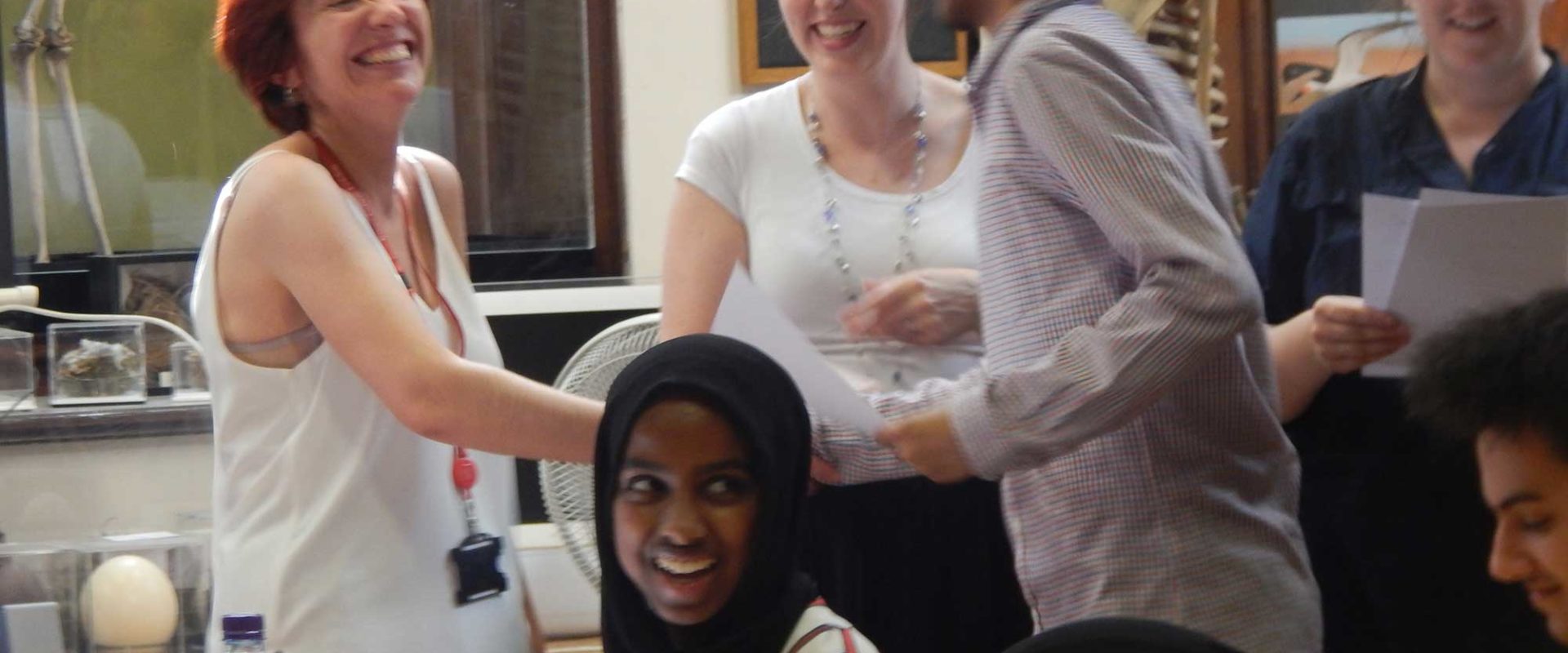
Farzana learnt about glial cell research during her two-week placement under the supervision of Angshu Angbohang, in the group of Professor Astrid Limb.
During my two weeks at the UCL Institute of Ophthalmology I’ve learnt about the human glial cell, a type of stem cell, that is able to regenerate into retinal neurons. This is important because the retinal neurons are photoreceptors at the back of the human retina which enables you to sense light and therefore allows you to see.
Scientists have discovered that the Muller glial cells in zebrafish have the ability to regenerate the retina neurons multiple times once they are damaged. This ultimately intrigues us to find out why and how they are able to do that and if one day a human may be able to do the same.These are the steps that I have taken during my placement, they all link together therefore it was important to do each step carefully as any mistake could of had an effect onto the final result:
Cell culture
Firstly I saw two Petri dishes that had cells inside of them, however one dish was confluent and the other dish had cells that were sparsely separated. We used the confluent dish which had to be vacuumed as there was solution around the cells, this gave us the cells we needed and nothing else. Furthermore, the cells were attached to the dish, due to this we had to use a solution called triple E as this helped detach the cells from the surface of the Petri dish. We then placed the dish into an incubator at 37 degrees Celsius which was followed by centrifuging the cells. Five minutes later the cells were at the bottom of the dish, we then removed the triple E by aspirating it and adding 5ml of medium (which provide nutrients to the cell). Finally we stained the cells using methyl blue.
RNA Extraction
We then extracted RNA from the cells we cultured. I removed medium first and then added the triple E solution as this helps detach the cells from the dish. We then centrifuged the cells which left a cell plate present (a white disk at the bottom). We then removed the triple E solution to give us the cells that we needed to extract the RNA.
We added lysis buffer (one micro litre) and beta-mercaptoethanol (a corrosive alcohol), we mixed them together and added 350 micro litres into control and treatment tubes. After that we used a syringe to help move the solution up and down, to help the breakdown with the lysis buffer to go faster. I centrifuged the cells again and then transfered the solution into an eppendorf tube.
Reverse Transcription
Reverse transcription is when the RNA turns into cDNA by using mRNA, primers, DTT and a reverse transcriptase enzyme (such as “Superscript”)
PCR
The polymerase chain reaction (PCR) is to help the cDNA to multiply rapidly and the forward and reverse primers bind to each strand. We needed cDNA, primer, water and Gotaq (a green solution that contained everything). We use cDNA because we know how long it lasts, however a normal DNA can affect the results.
 The primers use to test for photoreceptor are:
1) NR2E3
2) Recoverin
3) IRBP
4) Betactin
This was done because we needed space for the reverse transcription solution (mRNA, primer, DTT and superscript). To do this we need agarose gel, solution buffer and red gel (something that allows you to see the band).
Analysis


